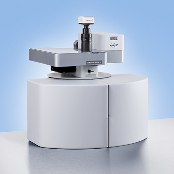
SENTERRA II Compact Raman Microscope from Bruker Optics
The SENTERRA II defines a new level of spectroscopic performance and user friendliness in the cla……...


The Thermo Scientific Volumescope 2 Scanning Electron Microscope (SEM) is an advanced serial block-face imaging system. The device features user-friendly technology to maintain control over research and safeguard a variety of potential samples with tried-and-tested solutions for each stage of the acquisition process.
Block Face Imaging
Understanding the intricate three-dimensional structure of biological systems and soft materials is essential for understanding the relationship between structure and function. For automated, three-dimensional (3D) reconstruction of large tissue volumes, serial block-face SEM (SBF-SEM) combines in situ sectioning and imaging of plastic-embedded tissue blocks within the SEM vacuum chamber.
The minimum section thickness once limited axial resolution, but truly isotropic resolution is now possible with the addition of multi-energy deconvolution.
3D SEM
After the block face is sectioned in situ using a diamond knife, the newly exposed tissue is photographed numerous times at progressively higher accelerating voltages. These images are then used in a deconvolution algorithm to derive several optical subsurface layers, forming a 3D subset. By repeating this cycle, the Volumescope 2 SEM provides isotropic datasets with 10 nm Z-resolution.
Mouse brain sample imaged on the Volumescope 2 SEM. Fine features such as vesicles and synaptic clefts can be observed. Image Credit: Dr Gabor Nyiri, Institute of Experimental Medicine, Budapest, Hungary

Volume reconstruction of a mouse heart muscle. Data acquired by a combination of physical and virtual slicing in high vacuum mode. Volume = 16×15×11 μm3 with a nominal resolution of 5×5×10 nm. Specimen courtesy: Dr Madesh Muniswamy, Temple University, USA. Image Credit: Thermo Fisher Scientific – Electron Microscopy Solutions
Polymer Microstructures
High-resolution imaging in 3D is imperativewhen trying to understand the properties and microstructure of polymer materials. The Volumescope 2 SEM combinesin situsectioning and imaging of polymers within the SEM vacuum chamber in a fully automated fashion to allow for the reconstruction of large volumes with truly isotropic 3D resolution.
Visualizing a sizable volume at these resolutions is essential to show how areas with various microstructures impact the material’s overall properties. To improve performance forecasts for new filter designs, the Volumescope 2 SEM, for instance, can recreate a large representative volume of a filtration membrane, providing accurate transport properties.

Serial block-face 3D imaging of a polymer blend sample. Here, the 3D volume reconstruction of the complete dataset (122 μm × 122 μm × 10 μm) is shown, with a segmented region inset revealing the particle distribution in the matrix. Sample courtesy of Dr. Armin Zankel (FELMI-ZFE Graz, Austria). Image Credit: Thermo Fisher Scientific – Electron Microscopy Solutions
Key Features
Easy-to-Use Technology
Easy-to-use technology allows users to stay in control of their experiments.Reuse jobs and system settings and select multiple regions of interest during job acquisition.
Protect Valuable Samples
Protect valuable samples with tested solutionsat every acquisition step:debris trap and swipe features ensure sample quality; low vacuum detector enables imaging of highly charged samples.
Visualize and Navigate During the Acquisition
Visualize and navigate during acquisitionwith Thermo Scientific Amira Live Tracker Software to optimize/control your outcome/Automate large 3D volume acquisitions as well as reconstructions.
Easy and Fast Mounting Microtome Exchange
Easy and fast mounting microtome exchangefor normal SEM operation or automated array tomography with the addition ofThermo Scientific Maps Software.
Specifications
Source: Thermo Fisher Scientific – Electron Microscopy Solutions
| . | . |
|---|---|
| Electron optics |
High-stability Schottky field emission gun Compound final lens: a combined electrostatic, field-free magnetic and immersion magnetic objective lens |
| Source lifetime |
24 months |
| Automation |
|
| Stage |
Double stage scanning deflection |
| Electron beam |
In serial block-face imaging SEM: 50 pA – 3.2 nA In serial block-face imaging SEM: 500 V – 6 kV 0.8 nm at 30 keV STEM 0.7 nm at 15 keV 1.0 nm at 1 keV |
| Chamber |
|
| Detectors |
T1 segmented lower in-lens detector VS-DBS: LoVac lens-mounted BSED |
| Vacuum system |
|
| Sample holders |
|
The SENTERRA II defines a new level of spectroscopic performance and user friendliness in the cla……...

As one of the world’s largest processor and supplier of natural and synthetic diamond powde……...

4D Biomaterial's mission is to commercialize a new class of materials –liquid resins t……...
![[db:国别翻译]德国Kistler Maschinen- und Anlagenbau GmbH](/wp-content/uploads/25upplier25/full_site_logo.png)
奇斯特勒机械公司是一家历史悠久的家族企业,致力于为焊接行业(尤其是管道焊接和切割)设计和制造焊接……...
/9-9-I-9M-9M-9-9A-9-9G-9G-9E-1/ Spotwelding Consultants,Inc。成立于1983年,为我们的客户提供了……...

CDET OMEGA is a shock tube based initiating device used for true bottom initiation of the bore ho……...

Item North America is a premier provider of precision ball screws for use as a drive mechanism in linear slides, multi-axis motion and complex material handling devices. item ball screws feature hi……
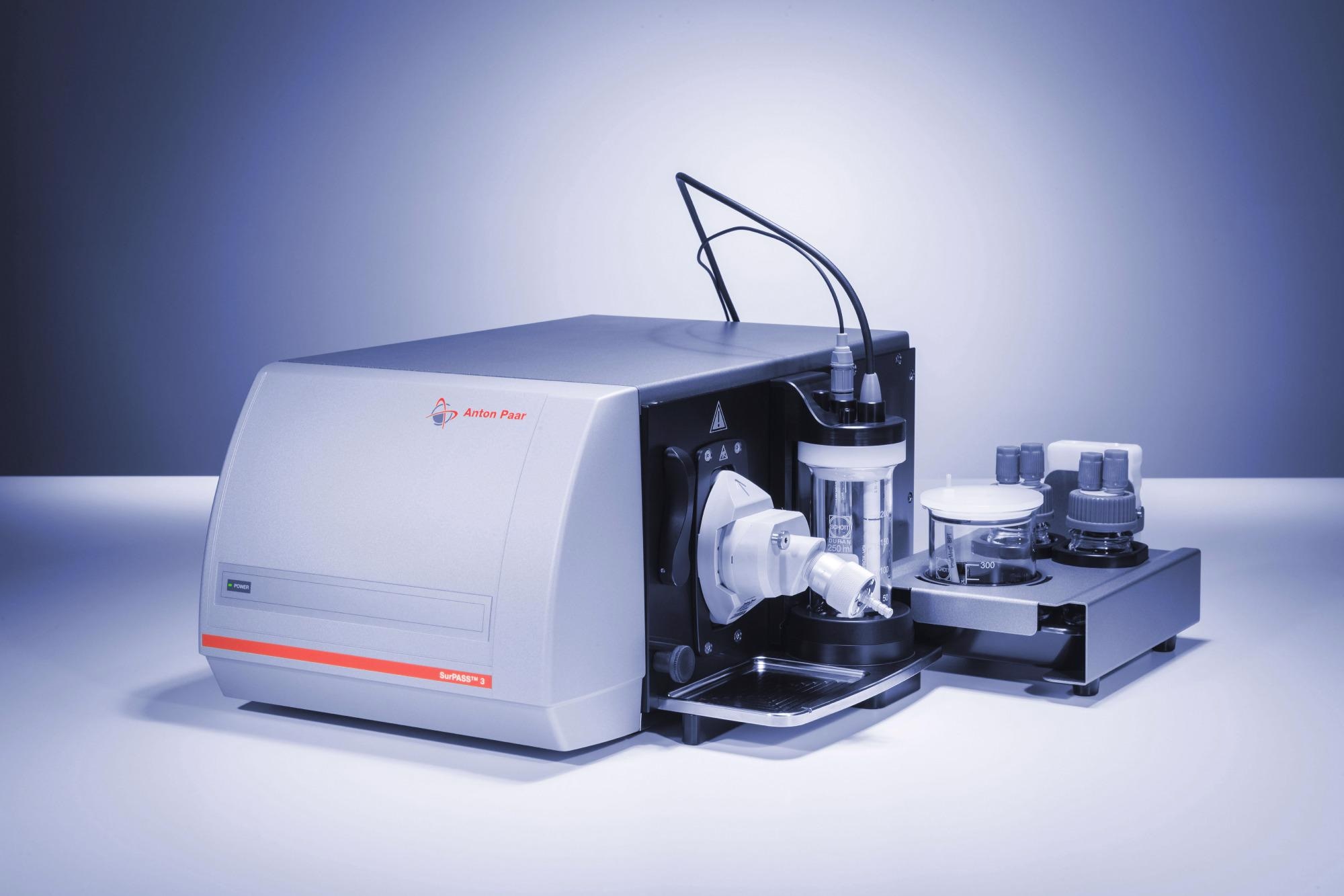
SurPASS™ 3 is capable of measuring the zeta potential over an extensive range of materials and allows the analysis of varied surface properties and their changes. The zeta potential explains ……

AHP Materials is a specialty materials company focused on the manufacturing and selling of high purity specialty metals and compounds. AHP concentrates mainly on Antimony, Cadmium Sulfide, Telluriu……